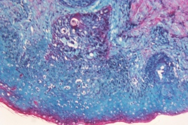
Odland bodies - Small, granular, membrane-bound vacuoles found in the cytoplasm of skin keratinocytes. Also known as keratinosomes or lamellar bodies, they are derived from the Golgi apparatus and are associated with the storage and release of lipid precursors. The figure shows the synthesis of epidermal lipids where 1=the odland bodies, 2=stratum granulosum cells, 3=exocytosis, 4=bilayer lipid membrane and 5=the cells of the stratum corneum
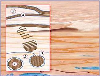
Odland bodies
|
Oken bodies
|
Oken bodies - Primitive or primordial kidneys of the embryo (mesonephron) formerly called Wolffian bodies
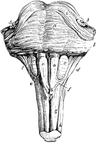
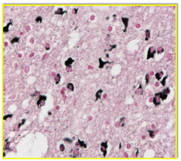
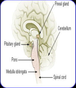
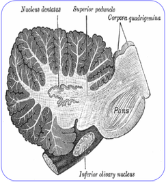
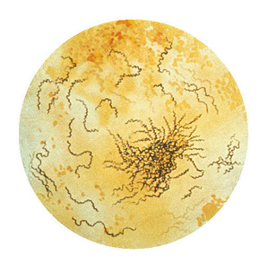
Olivary bodies - Rounded elevations lateral to the upper part of each pyramid of the medulla oblongata. Known simply as the olives, they are formed by the olivary nucleus just beneath its surface and are linked to the pons and cerebellum. The figure shows: a, anterior pyramids; b, their decussation; c, olivary bodies; d, restiform bodies; e, arciform fibres; f, fibres passing from the anterior column of the cord to the cerebellum; g, anterior column of the spinal cord; h, lateral column; p, pons Varolii; i, its upper fibres; 5, roots of the fifth pair of nerves
Olivary bodies - Rounded elevations lateral to the upper part of each pyramid of the medulla oblongata. Known simply as the olives, they are formed by the olivary nucleus just beneath its surface and are linked to the pons and cerebellum. The figure shows: a, anterior pyramids; b, their decussation; c, olivary bodies; d, restiform bodies; e, arciform fibres; f, fibres passing from the anterior column of the cord to the cerebellum; g, anterior column of the spinal cord; h, lateral column; p, pons Varolii; i, its upper fibres; 5, roots of the fifth pair of nerves
Olivary bodies (labelled 'c')
|
Orbital fat body
|
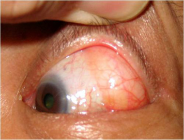
Orbital fat bodies - Fatty masses in the orbit which contribute to support of the eyeball. They are also known as orbital fat pads or corpus adiposum orbitae. With increasing age (or because of genetic disposition), the eyeball can descend and reduce the space between it and the floor of the orbit. This will result in forward projection of the orbital fat bodies which can create hernias
Orphan bodies - see Nuclear bodies
Oryzoid bodies - see Rice bodies
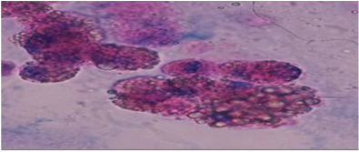
Oval fat bodies - These are degenerating renal tubular epithelial cells or macrophages containing abundant lipid. They appear as grape-like clusters of variable size and are highly refractile, exhibiting Maltese cross birefringence under polarized light. They are associated with lipiduria and are found in the urine of patients with diseases such as glomerulonephritis and nephrotic syndrome
Orphan bodies - see Nuclear bodies
Oryzoid bodies - see Rice bodies
Oval fat bodies - These are degenerating renal tubular epithelial cells or macrophages containing abundant lipid. They appear as grape-like clusters of variable size and are highly refractile, exhibiting Maltese cross birefringence under polarized light. They are associated with lipiduria and are found in the urine of patients with diseases such as glomerulonephritis and nephrotic syndrome
Oval fat bodies
|
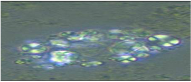
Oval fat bodies showing Maltese cross birefringence
|
Owl's eye bodies - These are usually associated with the owl's eye inclusion bodies found in tissues infected with the cytomegalovirus infection, a disease which can cause multiple organ dysfunction. However, the owl's eye appearance may also be seen in nuclei of Reed-Sternberg cells in patients with Hodgkin’s lymphoma
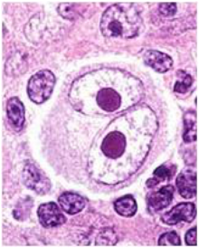
Owl's eye bodies
|
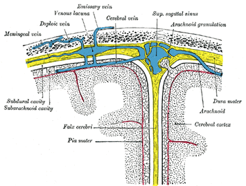
Pacchionian bodies (arachnoid granulations)
|
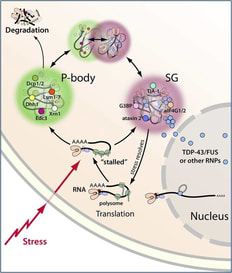
P bodies - see Processing bodies
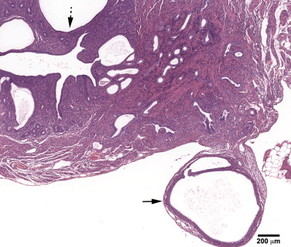
Pacchionian bodies - Enlarged fleshy processes or villi on the outer surface of the dura mater of the brain, also called arachnoid villi or arachnoid granulations. They act as one-way valves for the flow of CSF from the subarachnoid space into the bloodstream. In adults and the elderly, the villi become larger and are called pacchionian bodies
Pacini bodies - see Vater-Pacini bodies
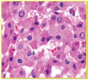
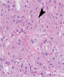
Pale bodies - Pale hyaline inclusion bodies found in the substantia nigra of the brain in patients with Parkinson’s disease. These bodies are membrane-bound, contain granular, electron dense material and are thought to be closely related to rough endoplasmic reticulum. They have also been found in hepatocellular carcinoma
Pale bodies
|
Pampiniform body (epoophoron)
|
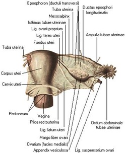
Pampiniform bodies - Rudimentary organs homologous with the male epididymis that lie in the broad ligament of the uterus next to the ovary and fallopian tube. Also known as the epoophoron, parovarium and organ of Rosenmuller
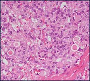
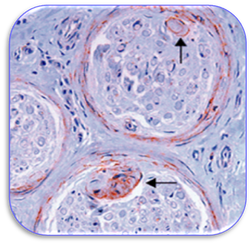
Papillary mesenchymal bodies - These are aggregations of fibroblasts that represent abortive attempts to form the papillary mesenchyme responsible for hair induction. These bodies are generally associated with trichoepithelioma, one of the tumours of the follicular epithelium. The presence of them is reliable in differentiating between trichoepithelioma and basal cell carcinomas
Papillary mesenchymal bodies - These are aggregations of fibroblasts that represent abortive attempts to form the papillary mesenchyme responsible for hair induction. These bodies are generally associated with trichoepithelioma, one of the tumours of the follicular epithelium. The presence of them is reliable in differentiating between trichoepithelioma and basal cell carcinomas
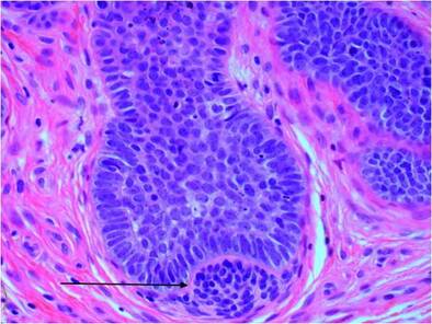
Papillary mesenchymal body (arrowed)
|
Pappenheimer bodies
|
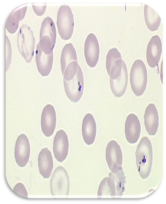
Pappenheimer bodies - Small irregular dark staining granules of iron formed by phagosomes and found in red blood cells in patients with anaemia
Papp-Lantos bodies - Cytoplasmic inclusion bodies found in glial cells of the brain in multiple-system atrophy (MSA), a degenerative neurological disorder. MSA causes cell loss and gliosis and proliferation of astrocytes in damaged areas of the central nervous system. The presence of these inclusions in the movement, balance, and automatic-control centres of the brain are the defining histological hallmark of MSA
Para aortic bodies – see Zuckerkandl bodies
Papp-Lantos bodies impregnated with silver
|
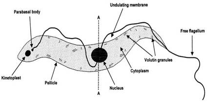
Parabasal body
|
Parabasal bodies - Cytoplasmic bodies closely associated with the nuclei, kinetoplasts and basal bodies in certain parasitic flagellate protozoa. They are usually connected to the basal bodies by a fibril or thread, which together are known as the parabasal apparatus (also see Basal bodies)
Paramural bodies – Membranous structures of plant cells located between the cell wall and plasma membrane. The structure may also be termed a lomasome (if it contains an internal membrane) or a plasmalemmasome if it is absent. The figure shows an electron micrograph of paramural bodies (arrowed) located between the endoplasmic reticulum (ER) and cell wall (CW). Also see multivesicular and vesicle-like bodies
Paramural bodies – Membranous structures of plant cells located between the cell wall and plasma membrane. The structure may also be termed a lomasome (if it contains an internal membrane) or a plasmalemmasome if it is absent. The figure shows an electron micrograph of paramural bodies (arrowed) located between the endoplasmic reticulum (ER) and cell wall (CW). Also see multivesicular and vesicle-like bodies
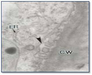
Paramural bodies
|
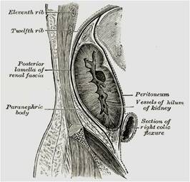
Paranephric body
|
Paranephric bodies - A collection of adipose tissue located superficial to the renal fascia. Also known as pararenal bodies
Paranuclear blue bodies - These inclusions are a feature of small cell carcinomas (SCC) in air dried, cytology smears stained with Romanowsky type stains. Their presence helps to differentiate SCC from non-small cell or poorly differentiated carcinomas in fine needle aspirates
Paranuclear blue bodies - These inclusions are a feature of small cell carcinomas (SCC) in air dried, cytology smears stained with Romanowsky type stains. Their presence helps to differentiate SCC from non-small cell or poorly differentiated carcinomas in fine needle aspirates
Paranuclear blue bodies
|
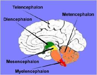
The diencephalon - see Paraphysial bodies
|
Paraphysial bodies - Organs that develop from the diencephalon in the forebrain of lower vertebrates. Also known as the paraphysis, these bodies may be present in the human embryo and foetus for a short time
Pararenal bodies - see Paranephric bodies
Paraterminal bodies - Slender vertical whitish bands near the anterior commissure of the brain. Also called the paraterminal gyrus and the peduncle of corpus callosum
Pararenal bodies - see Paranephric bodies
Paraterminal bodies - Slender vertical whitish bands near the anterior commissure of the brain. Also called the paraterminal gyrus and the peduncle of corpus callosum
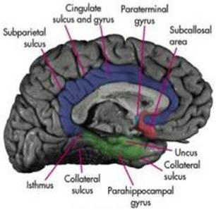
Paraterminal body or gyrus
|
Parietal body (arrowed)
|
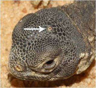
Parietal bodies - Eyelike structures on the dorsal aspect of the head of certain lower vertebrates. A modification of the parapineal organ, they are also termed epiphysial eyes and act as photoreceptors which enable response to darkness and light
Paschen bodies - Cellular inclusion bodies (usually in hepatocytes of the liver) found in patients with variola and vaccinia infections. It is not known whether the bodies are the infective agents or merely mechanical carriers of the virus
PcG (polycomb) bodies - see Nuclear bodies
Paschen bodies - Cellular inclusion bodies (usually in hepatocytes of the liver) found in patients with variola and vaccinia infections. It is not known whether the bodies are the infective agents or merely mechanical carriers of the virus
PcG (polycomb) bodies - see Nuclear bodies
Paschen bodies
Perineal bodies - Pyramidal fibromuscular mass in the middle line of the perineum at the junction between the urogenital triangle and the anal triangle. Also known as the central tendon of perineum, it is found in both males and females. In males, it is found between the bulb of penis and the anus; in females, is found between the vagina and anus. The perineal body is essential for the integrity of the pelvic floor, particularly in females. Its rupture during delivery leads to widening of the gap between the anterior free borders of levator ani muscle of both sides, thus predisposing the woman to prolapse of the uterus, rectum, or even the urinary bladder
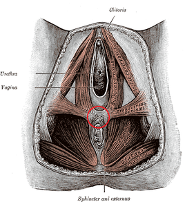
Perineal body (circled in red)
|
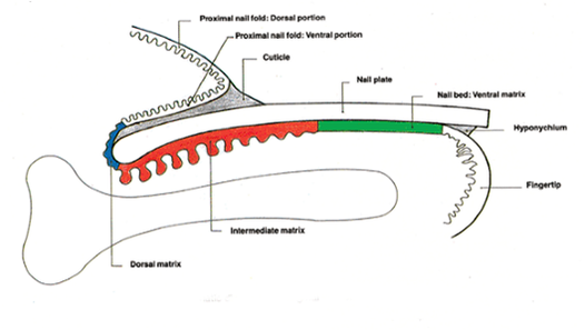
Diagram of fingertip showing nail plate (see Pertinax bodies)
|
Pertinax bodies - These are acidophil masses found in the nail plate and are remnants of the nuclei of keratinocytes. Known as pertinax bodies of Lewis and Montgomery, they are often associated with the nail changes that are common in the elderly
Pheochrome bodies - see Chromaffin bodies
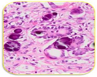
Pick bodies - Homogenous, round or ovoid structures found in histiocytes . They are strongly argyrophilic and are found in Niemann-Pick disease, a genetic disorder characterized by the accumulation of sphingomyelin. The disease is a relatively rare, degenerative brain illness that causes dementia. The symptoms are so variable that it overlaps with Alzheimer’s disease, though the presence of Pick bodies helps confirm the diagnosis
Pheochrome bodies - see Chromaffin bodies
Pick bodies - Homogenous, round or ovoid structures found in histiocytes . They are strongly argyrophilic and are found in Niemann-Pick disease, a genetic disorder characterized by the accumulation of sphingomyelin. The disease is a relatively rare, degenerative brain illness that causes dementia. The symptoms are so variable that it overlaps with Alzheimer’s disease, though the presence of Pick bodies helps confirm the diagnosis
Pick bodies
|
Pineal body (gland)
|
Pineal bodies - A small conical structure attached by a stalk to the posterior wall of the third ventricle in the centre of the brain. Also termed pineal glands or epiphysis cerebri, they secrete melatonin
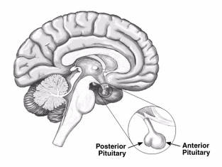
Pituitary bodies - This is another term for the pituitary glands that are located at the base of the brain. They are part of the endocrine system and rest in a small, bony cavity covered by a fold. The pituitary gland secretes nine hormones that regulate homeostasis
Pituitary bodies - This is another term for the pituitary glands that are located at the base of the brain. They are part of the endocrine system and rest in a small, bony cavity covered by a fold. The pituitary gland secretes nine hormones that regulate homeostasis
Pituitary body
|
Polar bodies
|
Platelet dense bodies - see Dense bodies
PML (Promyelocytic leukaemia) bodies - see Nuclear bodies
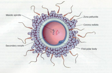
Polar bodies - A minute cell (sometimes termed the polar cell or globule) that separates from the ovum in the maturation of ordinary ova. The first polar body formed is usually the largest and often divides into two after separation. Each body removes maternal chromatin from the ovum to make room for the chromatin of the fertilizing sperm
Polycomb (PcG) bodies - see Nuclear bodies
Polyglucosan bodies – see Amyloid bodies
Postbranchial bodies - see Ultimobranchial bodies
Poulsen-Christoffersen bodies - These are damaged bile ducts that are frequently associated with viral hepatitis. Commonly known as Poulsen-Christoffersen lesions, the damaged epithelium of the bile ducts is more widespread in hepatitis C infection. These ducts are characterized by vacuolation of the cells with overlapping of the nuclei and cytoplasmic eosinophilia
PML (Promyelocytic leukaemia) bodies - see Nuclear bodies
Polar bodies - A minute cell (sometimes termed the polar cell or globule) that separates from the ovum in the maturation of ordinary ova. The first polar body formed is usually the largest and often divides into two after separation. Each body removes maternal chromatin from the ovum to make room for the chromatin of the fertilizing sperm
Polycomb (PcG) bodies - see Nuclear bodies
Polyglucosan bodies – see Amyloid bodies
Postbranchial bodies - see Ultimobranchial bodies
Poulsen-Christoffersen bodies - These are damaged bile ducts that are frequently associated with viral hepatitis. Commonly known as Poulsen-Christoffersen lesions, the damaged epithelium of the bile ducts is more widespread in hepatitis C infection. These ducts are characterized by vacuolation of the cells with overlapping of the nuclei and cytoplasmic eosinophilia
Poulsen-Christoffersen lesion
|
Location of the Processing (P) body
|
Processing bodies - Commonly known as P bodies, these are distinct foci within the cytoplasm of the eukaryotic cell. They contain many enzymes that are involved in the turnover of messenger RNA. These bodies play essential roles in the general decay of mRNA although not all mRNAs which enter P bodies are degraded. P bodies are sometimes referred to as GW bodies (see separate entry for GW bodies) and are also associated with cytoplasmic U bodies (see U bodies)
Promyelocytic leukaemia (PML) bodies - see Nuclear bodies
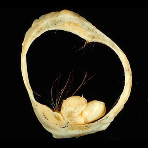
Protocorm-like bodies - In plant biology, these are small swollen tubers that consist of undifferentiated cells that have the ability to produce shoots in vitro. Commonly known as PLBs, they are involved in the propagation of plants, particularly orchids. The image shows the development of secondary PLBs from a single protocorm
Protocorm-like bodies - In plant biology, these are small swollen tubers that consist of undifferentiated cells that have the ability to produce shoots in vitro. Commonly known as PLBs, they are involved in the propagation of plants, particularly orchids. The image shows the development of secondary PLBs from a single protocorm
Protocorm-like bodies (PLBs)
|
Skin infected with Variola (see Prowazek bodies)
|
Prowazek bodies - Tiny, ovoid, granular bodies (frequently in pairs) and seen in the cytoplasm of cutaneous squamous cells. They occur in humans and animals infected with the variola (smallpox) or vaccinia virus
Psammoma bodies - Gritty, laminated, sand-like particles in various stages of hyaline change. Sometimes termed sand bodies, corpora arenacea and calcospherite, they are usually diagnostic of papillary carcinoma. These bodies are typically found in cancer of the ovary but may also be found in papillary carcinoma of the thyroid (also see Pseudopsammoma bodies)
Psammoma bodies - Gritty, laminated, sand-like particles in various stages of hyaline change. Sometimes termed sand bodies, corpora arenacea and calcospherite, they are usually diagnostic of papillary carcinoma. These bodies are typically found in cancer of the ovary but may also be found in papillary carcinoma of the thyroid (also see Pseudopsammoma bodies)
Psammoma bodies
|
Pseudoasbestos body
|
Pseudoasbestos bodies - Structures that form as a result of deposition of ferroproteins on inhaled material such as coal, silica, graphite and talc. This excludes bodies that result from the inhalation of asbestos fibres (also see Asbestos bodies)
Pseudopsammoma bodies - Eosinophilic, hyaline inclusion bodies often seen in meningiomas. They are PAS and CEA positive and may often have a granular appearance (also see Psammoma bodies)
Pseudopsammoma bodies - Eosinophilic, hyaline inclusion bodies often seen in meningiomas. They are PAS and CEA positive and may often have a granular appearance (also see Psammoma bodies)
Pseudopsammoma bodies
|
Pustulo-ovoid body (arrowed)
|
Pulmonary blue bodies - see Blue bodies
Pustulo-ovoid bodies - Round eosinophilic granules surrounded by a clear halo and found in granular cell tumours. Known as pustulo-ovoid bodies of Milian, they represent lysosomes and give the appearance of large granules that have partially detached from the adjacent cytoplasm. Pustulo-ovoid bodies are an easily recognizable component of these benign neural tumours
Quadrigeminal bodies - Two pairs of lobes found on the dorsal side of the midbrain. Known as corpora quadrigemina, they are the reflex centres for sight and hearing and are respectively named the inferior and superior colliculus. The anterior pair are called the nates and the posterior ones the testes
Quadregeminal body
|
Raspberry bodies
|
Raspberry bodies – These extracellular hyaline globules are characteristic of clear cell carcinoma of the ovary. They are believed to be the result of excessive formation of a basement membrane-like (collagen) substance. Cytologically diagnostic in ascitic fluids, they stain pink to purplish-red with Giemsa
Reducing bodies - Characteristic inclusion bodies found in affected muscle fibres in the rare muscle disorder known as reducing body myopathy. During infancy or early childhood, the disease exhibits rapidly progressive muscle weakness leading to death within the first five years of life
Reducing bodies - Characteristic inclusion bodies found in affected muscle fibres in the rare muscle disorder known as reducing body myopathy. During infancy or early childhood, the disease exhibits rapidly progressive muscle weakness leading to death within the first five years of life
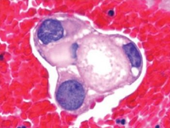
Reducing bodies stained dark with Gomori trichrome
|
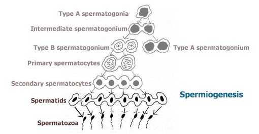
Spermiogenesis (see Regnaud body)
|
Regnaud bodies - Residual masses of cytoplasm cast off during the process of spermiogenesis (see figure). They consist of fine granules, lipid droplets and degenerating organelles. Spermiogenesis is the final stage of spermatogenesis which sees the maturation of spermatids into mature and motile spermatazoa
Reilly bodies – see Alder-Reilly bodies
Reinke bodies - Rod-like, cytoplasmic inclusions found in Leydig cells of the testes. Also known as Reinke crystals, they are seen in Leydig cell tumours and can be visualized with stains such as Giemsa, PAS and trichrome
Reinke bodies - Rod-like, cytoplasmic inclusions found in Leydig cells of the testes. Also known as Reinke crystals, they are seen in Leydig cell tumours and can be visualized with stains such as Giemsa, PAS and trichrome
Reinke bodies
|
Renaut bodies
|
Renaut bodies - Subperineurial structures comprised of loosely arranged and randomly oriented collagen fibres in a fine fibrillary material. They are seen in normal nerve as well as in certain pathologic states
Residual bodies - Vesicles containing undigestable material found in lysosomes. The material is called lipofuchsin and is often referred to as ‘wear and tear’ pigment. Residual bodies may be secreted by macrophages or become lipofuchsin which can remain in the cells indefinitely. Cells that live longer (such as neurones and muscle cells) have higher concentrations of lipofuchsin
Residual bodies - Vesicles containing undigestable material found in lysosomes. The material is called lipofuchsin and is often referred to as ‘wear and tear’ pigment. Residual bodies may be secreted by macrophages or become lipofuchsin which can remain in the cells indefinitely. Cells that live longer (such as neurones and muscle cells) have higher concentrations of lipofuchsin
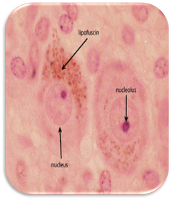
Residual bodies
|
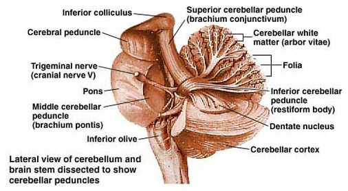
Restiform body
|
Restiform bodies - These are a subdivision of the inferior cerebellar peduncle, (which comprise both the juxtarestiform and restiform bodies) and carry both afferent and efferent nerve fibres. The bodies have a role in coordinating balance and eye movements (also see Juxtarestiform bodies)
Reticulate bodies – Chlamydia may be found in the form of an elementary and reticulate (initial) bodies. The reticulate body is a spherical, intracytoplasmic form highly involved in the process of replication and growth (also see Elementary and Halberstaedter-von Prowazek bodies)
Reticulate bodies – Chlamydia may be found in the form of an elementary and reticulate (initial) bodies. The reticulate body is a spherical, intracytoplasmic form highly involved in the process of replication and growth (also see Elementary and Halberstaedter-von Prowazek bodies)
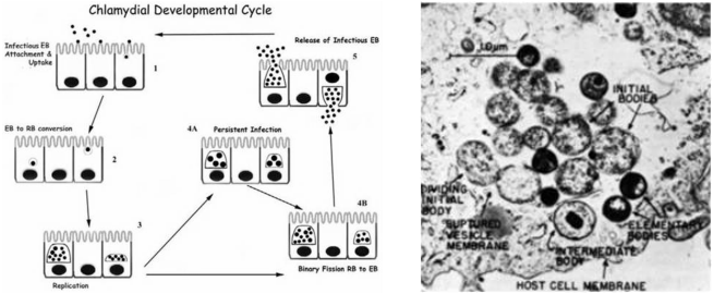
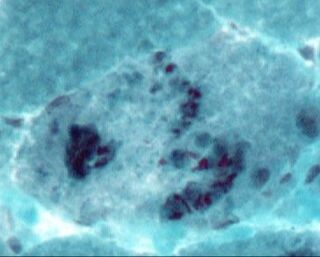
Reticulate bodies (RB)
Retzius bodies - Protoplasmic masses in the ear containing pigment granules. They are found at the lower end of the hair cells of the acoustic papilla (see image), a prominent ridge of highly specialised epithelium in the floor of the cochlear duct. They contain one inner row and three outer rows of hair cells and are associated with Hensen bodies
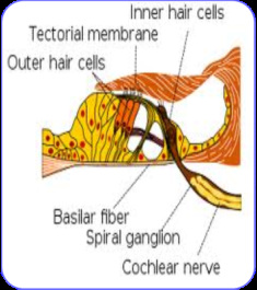
Acoustic papilla (see Retzius bodies)
|
Rice bodies
|
Rice bodies - These are intra-articular loose bodies macroscopically similar to grains of rice and most commonly diagnostic of rheumatoid arthritis. Sometimes termed Oryzoid bodies, they represent particles containing collagen, fibrinogen, fibrin, blood cells and amorphous material. Removal of them is usually accompanied by clinical improvement and a reduction of synovitis (also see Melon seed bodies)
Rod bodies - see Nemaline bodies
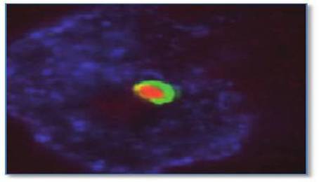
Rodlet bodies - Intranuclear inclusions predominantly seen in neurones in normal and neoplastic neuronal disease. They are believed to consist of small clusters of aligned filaments which increase in number as neuronal differentiation progresses. The image below shows double immunostaining of a neuronal intranuclear rodlet body with PML protein (green) and tubulin (red). Rodlet bodies have also been documented in dermal papillae (the cells which exert controlling influences on hair growth) and in animal systems
Rod bodies - see Nemaline bodies
Rodlet bodies - Intranuclear inclusions predominantly seen in neurones in normal and neoplastic neuronal disease. They are believed to consist of small clusters of aligned filaments which increase in number as neuronal differentiation progresses. The image below shows double immunostaining of a neuronal intranuclear rodlet body with PML protein (green) and tubulin (red). Rodlet bodies have also been documented in dermal papillae (the cells which exert controlling influences on hair growth) and in animal systems
Rodlet body - see text above
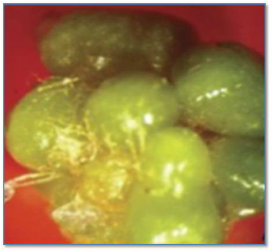
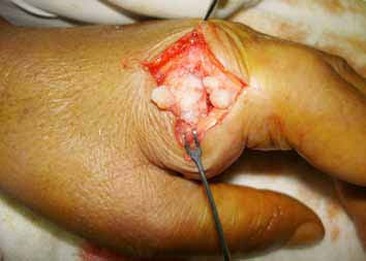
Rokitansky bodies - These are found in benign cystic teratomas and have the yellowish appearance of adipose tissue on CT scan. Sebaceous material and teeth may be seen inside the nodules
Rokitansky bodies
|
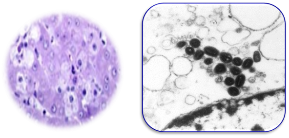
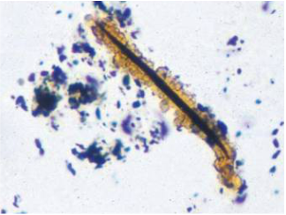
Treponema pallidum (see Ross bodies)
|
Ross bodies - Spherical copper-coloured bodies sometimes have amoeboid movement. They are found in the blood and tissue fluids of patients with syphilis, caused by the Gram negative spirochaete Treponema pallidum (see image)
Rushton bodies - Intraepithelial, curvilinear and eosinophilic lamellar structures found in odontogenic cysts. They commonly occur in the oral cavity and appear as amorphous concretions that are consistently positive for calcium and iron
Rushton bodies - Intraepithelial, curvilinear and eosinophilic lamellar structures found in odontogenic cysts. They commonly occur in the oral cavity and appear as amorphous concretions that are consistently positive for calcium and iron
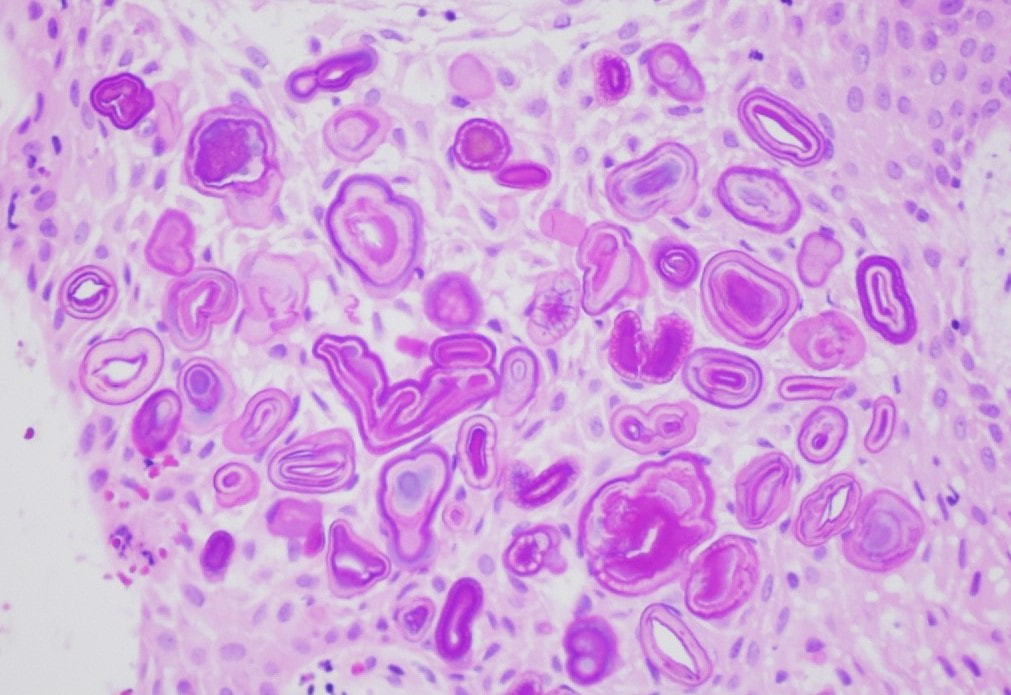
Rushton bodies
|
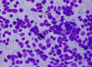
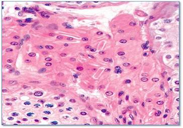
Russell bodies staining positive with fuchsin
|
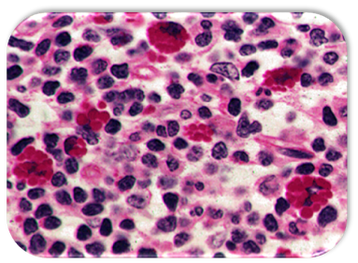
Russell bodies - Spherical cytoplasmic hyaline bodies in plasma cells associated with chronic inflammation and cancers such as myeloma and plasmacytoma. They are also known as Fuchsin bodies, Unna bodies and Russell-Krukenberg bodies and represent deposits of gamma globulin. The cells that contain these structures are known as Mott bodies or cells (see Mott bodies)
Russell-Krukenberg bodies – see Russell bodies
Russell-Krukenberg bodies – see Russell bodies