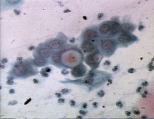
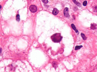
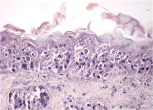
Kamino bodies - These are eosinophilic globoid bodies (KB) that are probably apoptotic lesional cells. They are found in spindle cell naevi but usually absent from malignant melanomas
Keratin bodies - see Fibrous bodies
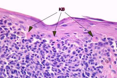
Kamino bodies
|

Ketone bodies
|
Ketone bodies - Ketone (or Acetone) bodies comprise acetone, acetoacetic acid and beta-hydroxybutyric acid. They are produced by the liver as a result of fat metabolism and are normally used as energy sources by tissues such as muscle. If supply of carbohydrate becomes limited, the blood level of ketone bodies rise. They may also be present in urine, giving it the characteristic odour of pear drops (ketosis)
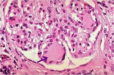
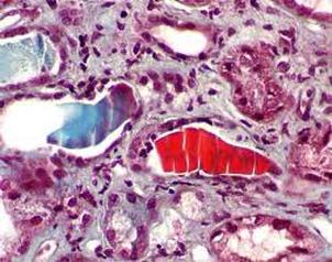
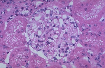
Kimmelstiel-Wilson bodies - Rounded areas of pink, hyaline material found in nodular glomerulosclerosis of the kidney. Termed the Kimmelstiel-Wilson syndrome, this renal disease is found in patients with diabetes and affects the network of tiny blood vessels in the glomerulus. Features of the disease include excessive protein in the urine, high blood pressure and progressively impaired kidney function
Kimmelstiel-Wilson bodies - Rounded areas of pink, hyaline material found in nodular glomerulosclerosis of the kidney. Termed the Kimmelstiel-Wilson syndrome, this renal disease is found in patients with diabetes and affects the network of tiny blood vessels in the glomerulus. Features of the disease include excessive protein in the urine, high blood pressure and progressively impaired kidney function
Kimmelstiel-Wilson bodies
Koch (or Koch's blue) bodies - These are schizonts of the protozoa Theileria parva, the causative agent of east coast fever of cattle, sheep and goats. The disease is transmitted by ticks and when the animals are infested with them, small unicellular bodies appear in the tissue
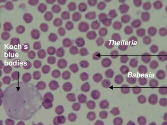
Koch's blue bodies
|
Kurloff bodies
|
Kurloff bodies - Large, slightly granular, cytoplasmic inclusions found in mononuclear leucocytes (Kurloff cells). The cells are unique to guinea pigs and have immune and natural killer characteristics. The bodies are composed of mucopolysaccharides and are PAS-positive
Lafora bodies - Cytoplasmic, basophilic and metachromatic inclusions having a concentric target like lamination. They are found in nerve cells but may also be found in the liver, muscle and skin of patients with Lafora body disease. The bodies are Alcian blue and PAS positive but resistant to diastase. Lafora body disease is a metabolic storage disease that causes progressive epilepsy in the late childhood. Clinical features include rapid intellectual decline and development of dementia
Lafora bodies - Cytoplasmic, basophilic and metachromatic inclusions having a concentric target like lamination. They are found in nerve cells but may also be found in the liver, muscle and skin of patients with Lafora body disease. The bodies are Alcian blue and PAS positive but resistant to diastase. Lafora body disease is a metabolic storage disease that causes progressive epilepsy in the late childhood. Clinical features include rapid intellectual decline and development of dementia
Lafora bodies
|
Lallemand bodies
|
Lallemand bodies - These are slightly irregular, rod-shaped or cylindrical bodies of proteinaceous material often seen in seminal fluid. Sometimes known as Trousseau-Lallemand bodies or Bence Jones cylinders, they are suggestive of multiple myeloma or macroglobulinaemia
Lamellar bodies - Concentric laminated, scroll-like whorls seen in alveolar spaces of lung. They are found in patients with pulmonary mucosa-associated lymphoid tissue (MALT) lymphomas and sclerosing haemangiomas of the lung. Lamellar bodies are acidic, surfactant-secreting organelles of lung epithelia. Also see Odland bodies
Lamellar bodies - Concentric laminated, scroll-like whorls seen in alveolar spaces of lung. They are found in patients with pulmonary mucosa-associated lymphoid tissue (MALT) lymphomas and sclerosing haemangiomas of the lung. Lamellar bodies are acidic, surfactant-secreting organelles of lung epithelia. Also see Odland bodies
Lamellar bodies
|
Leishman-Donovan bodies
|
Leishman-Donovan bodies - Small intracytoplasmic spheres found in the liver and spleen in patients with kala azar (Dum Dum fever) and leishmaniasis. The bodies are a non flagellated form of the parasitic protozoan Leishmania donovani and occur once the parasite has invaded the cells of the reticuloendothelial system
Levinthal-Coles-Lillie bodies - Cytoplasmic inclusion bodies found in macrophages of the lung in psittacosis, an infectious disease caused by Chlamydia psittaci having mild, flu-like symptoms. The infectious particles are called elementary bodies when they are shed from birds. They attach to and penetrate host cells and transform into non-infectious reticulate bodies
Levinthal-Coles-Lillie bodies - Cytoplasmic inclusion bodies found in macrophages of the lung in psittacosis, an infectious disease caused by Chlamydia psittaci having mild, flu-like symptoms. The infectious particles are called elementary bodies when they are shed from birds. They attach to and penetrate host cells and transform into non-infectious reticulate bodies
Levinthal-Coles-Lillie bodies
|
Lewy bodies
|
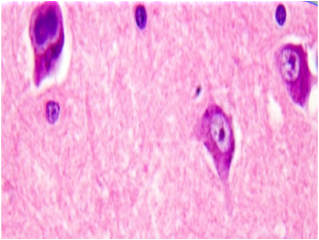
Lewy bodies - Round eosinophilic structures found in the cytoplasm of neurones. They may be single or multiple and are found in Dementia with Lewy bodies. They are also associated with both Parkinson's and Alzheimer’s disease. They are the result of altered neurofilament metabolism due to neuronal damage and stain with a variety of antibodies such as ubiquitin and neurofilament
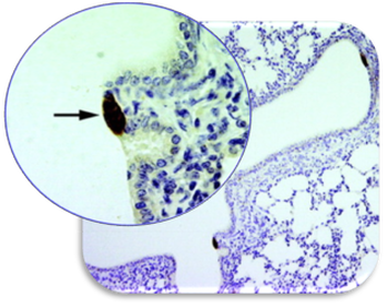
Lindner bodies - Elementary cytoplasmic structures found in inclusion conjunctivitis of the newborn (see image). The bacterial infection is caused by Chlamydia which infects the eyes of the baby during birth from an infected mother
Lindner Initial bodies – see Lindner bodies
Lindner bodies - Elementary cytoplasmic structures found in inclusion conjunctivitis of the newborn (see image). The bacterial infection is caused by Chlamydia which infects the eyes of the baby during birth from an infected mother
Lindner Initial bodies – see Lindner bodies
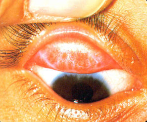
Conjunctivitis - see Lindner bodies
|
Lipschultz bodies
|
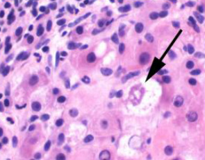
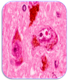
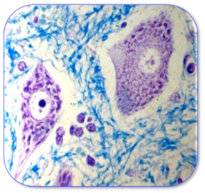
Lipschultz bodies - Intranuclear acidophilic inclusion bodies of cells found in ulcers of herpes simplex. The figure shows a multinucleated giant epithelial cell with intracytoplasmic basophilic inclusions, Lipschutz type 2 bodies. They are found during primary infection with herpes simplex virus. Herpes simplex virus type 1 is generally associated with oral and ocular infections while the type 2 virus is associated with genital infections
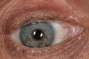
Lisch bodies - These are pigmented yellow-brown nodules that project from the surface of the iris in patients with type 1 neurofibromatosis. Also know as iris hamartomas, these bodies are aggregates of dendritic melanocytes and stain positive with S100 and vimentin. Lisch nodules are also found in Watson syndrome, an autosomal dominant condition characterised by pulmonary stenosis, macrocephaly and neurofibromas
Lisch bodies - These are pigmented yellow-brown nodules that project from the surface of the iris in patients with type 1 neurofibromatosis. Also know as iris hamartomas, these bodies are aggregates of dendritic melanocytes and stain positive with S100 and vimentin. Lisch nodules are also found in Watson syndrome, an autosomal dominant condition characterised by pulmonary stenosis, macrocephaly and neurofibromas
Lisch bodies
|
Loose bodies
|
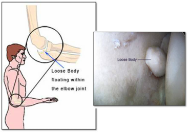
Loose bodies - Fragments of bone or cartilage that become detached into the joint space. These bodies may continue to grow by surface apposition and their centres may eventually become necrotic and calcify. Peritoneal loose bodies are masses that form from fat-filled pouches of the peritoneum (epiploic appendages) which may become detached and fibrosed. Often symptomless, they can cause compression of the of the bladder and bowel if they increase in size
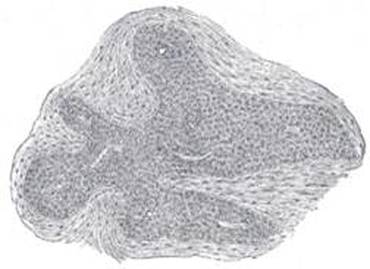
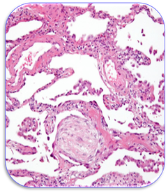
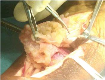
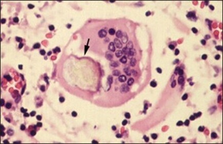
Luschka bodies - Also known as coccygeal bodies or glomus coccygeum, they are placed in front of, or immediately below, the tip of the coccyx. The bodies are innervated and correspond to a complex anastomosis between the sacral artery and vein, possibly having a haematopoietic function. The image shows a Lushka body nodule showing the epithelial and vascular patterns
Luschka bodies - Also known as coccygeal bodies or glomus coccygeum, they are placed in front of, or immediately below, the tip of the coccyx. The bodies are innervated and correspond to a complex anastomosis between the sacral artery and vein, possibly having a haematopoietic function. The image shows a Lushka body nodule showing the epithelial and vascular patterns
Luschka bodies
|
Luse bodies
|
Luse bodies - Parallel tropocollagen fibrils with a 120-130nm periodicity. They are found in rheumatoid disease and schwannoma. In rheumatoid connective tissue, detection of Luse bodies can occur with dysplastic and intracellular collagen
Luys bodies - This is another term for the lens-shaped subthalamic nuclei (corpus Luysii). They are found in the subthalamus of the brain and are part of the basal ganglia system
Luys bodies - This is another term for the lens-shaped subthalamic nuclei (corpus Luysii). They are found in the subthalamus of the brain and are part of the basal ganglia system
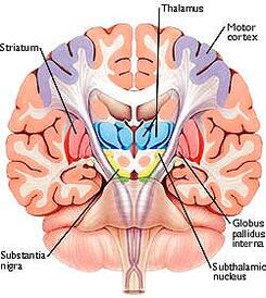
Luys bodies (subthalamic nuclei)
|
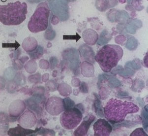
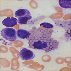
Lymphoglandular bodies in a large cell lymphoma
|
Lymphocytoid bodies - see Lymphoglandular bodies
Lymphoglandular bodies - These are fragments of cytoplasm that are well-organized from different types of cells in lymphoid tissue. In Giemsa stained imprints and smears, they appear as smooth, round, pale blue bodies with occasional projections. Also known as LGBs, Soderstrom or lymphocytoid bodies, they are indicative of tissues rich in lymphocytes and include lymphoid malignancies
Lymphoglandular bodies - These are fragments of cytoplasm that are well-organized from different types of cells in lymphoid tissue. In Giemsa stained imprints and smears, they appear as smooth, round, pale blue bodies with occasional projections. Also known as LGBs, Soderstrom or lymphocytoid bodies, they are indicative of tissues rich in lymphocytes and include lymphoid malignancies
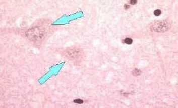 Lyssa bodies
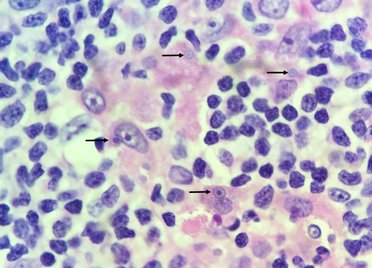
Lyssa bodies - Eosinophilic masses in
normal and degenerate neurones
resembling the Negri bodies found in
rabies. However, they are less sharply
defined and have less internal structure
(arrowed). Ultrastructural evidence
suggests that Lyssa bodies represent
Negri bodies without their histologically
recognizable inner bodies or cytoplasmic
inclusions
MacCallum bodies - Also known as MacCallum plaques, these are warty, irregular, mural lesions found in the endocardium of the heart in patients with rheumatic heart disease. In rheumatic fever, these bodies may be found in any layer of the heart and can cause different types of carditis
MacCallum bodies
|
Mallory bodies
|
Mallory bodies - Large accumulations of eosinophilic material in the cytoplasm of damaged liver cells. A perinuclear location is common and ring or twisted rope-like forms may occur. Also called alcoholic hyaline bodies, they are found in cirrhosis and alcohol-related diseases
Mallory-Denk bodies - Cytoplasmic, hyaline inclusion bodies of hepatocytes found in chronic hepatitis C and other liver diseases. They are believed to be another name for Mallory bodies. They are usually composed of keratins and other proteins such as ubiquitin and often occur with fatty change
Mallory-Denk bodies - Cytoplasmic, hyaline inclusion bodies of hepatocytes found in chronic hepatitis C and other liver diseases. They are believed to be another name for Mallory bodies. They are usually composed of keratins and other proteins such as ubiquitin and often occur with fatty change
Mallory Denk body
|
Malpighian bodies
|
Malpighian bodies - Part of a nephron of the kidney comprising the cup-shaped Bowman’s capsule and the glomerulus (the renal corpuscle). The Malpighian bodies, proximal and distal convoluted tubules lie in the cortex while the loop of Henle and main collecting ducts are in the medulla. Each Bowman’s capsule leads into a renal tubule which consist of descending and ascending tubules with the loop of Henle and collecting ducts
Mamillary bodies - These are a pair of small round masses located on the under surface of the brain. They belong to the limbic system and form part of the hypothalamus
Mamillary bodies - These are a pair of small round masses located on the under surface of the brain. They belong to the limbic system and form part of the hypothalamus
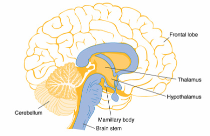
Mamillary body
|
Marchal bodies
|
Marchal bodies – Epithelial cell inclusion bodies observed in infectious ectromelia (mousepox). It is caused by the ectromelia virus and has only been seen in mouse colonies that are kept for research purposes. Mousepox is the only poxvirus to cause disease naturally in mice, producing skin lesions and generalized disease which can be fatal
Marinesco bodies - Circular, eosinophilic bodies mainly found in the nuclei of pigmented neurones of the substantia nigra. They are age related and thought to be associated with the onset of Parkinson’s disease
Marinesco bodies - Circular, eosinophilic bodies mainly found in the nuclei of pigmented neurones of the substantia nigra. They are age related and thought to be associated with the onset of Parkinson’s disease
Marinesco bodies
|
Masson bodies
|
Masson bodies - Connective tissue buds in the alveolar spaces and ducts of the lung. They are the hallmark of cryptogenic organising pneumonia (COP) and bronchiolitis obliterans organising pneumonia (BOOP). Masson bodies are polypoidal buds of proliferating fibroblasts and may be modified Aschoff bodies
Medlar bodies - see Sclerotic bodies
Medlar bodies - see Sclerotic bodies
|
Melamed-Wolinska bodies - These round cytoplasmic inclusions are found within degenerating benign or malignant urothelial cells. These globular bodies are usually red (eosinophilic bodies) but may also appear blue-green in colour. They have no diagnostic value in urine but may suggest a urothelial origin for malignant cells in effusions
|
 Melamed-Wolinska bodies Melamed-Wolinska bodies
|
Melon seed bodies - White or brownish oval bodies of fibrous synovial tissue that sometimes occur loose in numbers in the cavity of inflamed joints. Melon seed (and rice) bodies may be seen operatively in tuberculous tenosynovitis (also see Rice bodies)
Melon seed bodies
|
Michaelis-Gutmann bodies (arrowed)
|
Michaelis-Gutmann bodies - Round cytoplasmic inclusions having a lamellar appearance and which may contain calcium and iron. They often consist of round targetoid, bull's-eye calcospherites and are associated with malakoplakia, a chronic infection usually affecting the kidney. Malakoplakia is a rare chronic inflammatory process characterized by the collection of histiocytes with granular cytoplasm. The presence of Michaelis-Gutmann bodies represents the end result of imperfectly digested bacteria that were previously ingested by macrophages
Mid bodies – see Flemming bodies
Mikulicz bodies - These consist of large vacuolated, foamy histiocytes that are classically seen during the proliferative and chronic granulomatous phase of the disease rhinoscleroma. Commonly affecting the nasal cavity and nasopharynx, rhinoscleroma is caused by the organism Klebsiella rhinoscleromatis
Mikulicz bodies
|
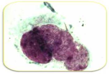
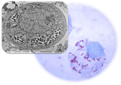
Miyagawa bodies (Chlamydia trachomatis)
|
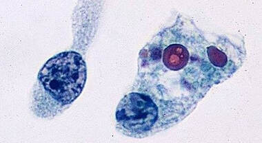
Miyagawa bodies - Elementary, intracytoplasmic microcolonies of Chlamydia trachomatis (see image). These are found in lymphogranuloma venereum (LGV), a sexually transmitted bacterial disease where the formation of buboes (lymph node swellings) is common
Molluscum bodies - see Henderson-Patterson bodies
Mooser bodies – see Neill-Mooser bodies
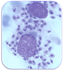
Mott bodies - Often termed Mott cells, these are plasma cells that have spherical inclusions (Russell bodies) packed in their cytoplasm. They are found in chronic inflammation and cancers such as myeloma and plasmacytoma (also see Russell bodies)
Molluscum bodies - see Henderson-Patterson bodies
Mooser bodies – see Neill-Mooser bodies
Mott bodies - Often termed Mott cells, these are plasma cells that have spherical inclusions (Russell bodies) packed in their cytoplasm. They are found in chronic inflammation and cancers such as myeloma and plasmacytoma (also see Russell bodies)
Mott bodies
|
Mulberry body (a) and a mulberry cell (b)
|
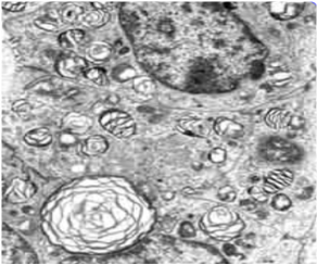
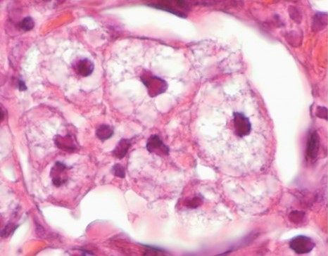
Mulberry bodies - Mulberry bodies are lamellar, spiral-shaped structures that are characteristic of Fabry disease, an inherited lysosomal storage disorder. This disease is caused by a deficiency of alpha galactosidase A which clinically manifests itself in adults with a decline in kidney function with eventual renal failure. A cardiac variant of Fabry disease has also been reported. Mulberry bodies are able to aggregate to form mulberry cells
Multilamellar bodies - Membrane-bound cellular organelles composed of concentric layers. They are found in numerous cell types but especially in the alveolar cells of the lung where they function in the storage and secretion of lipid. Multilamellar bodies are also found in the brain in tropical spastic paraparesis, a spinal cord infection caused by the human T-lymphotropic virus (HTLV)
Multilamellar bodies - Membrane-bound cellular organelles composed of concentric layers. They are found in numerous cell types but especially in the alveolar cells of the lung where they function in the storage and secretion of lipid. Multilamellar bodies are also found in the brain in tropical spastic paraparesis, a spinal cord infection caused by the human T-lymphotropic virus (HTLV)
Multilamellar bodies
|
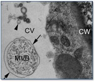
Multivesicular bodies
|
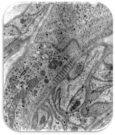
Multivesicular bodies - These are circular organelles that contain small vesicles formed by the membrane budding into the endosome. It is thought that they form paramural bodies by fusing with the plasma membrane. The figure shows a multivesicular body (MVB) in an epidermal cell with adjacent central vesicles (CV) and cell wall (CW). Also see paramural and vesicle-like bodies
Mushroom bodies - Also called corpora pedunculata, these are a pair of easily discernible structures in the brain of insects. They are also found in other arthropods (invertebrate animals having an exoskeleton) and some annelids (ringed worms). Comprising of thousands of densely-packed neurons, they are closely associated with the chemosensory system. The image shows a Drosophila brain where the mushroom bodies are highlighted in green
Mushroom bodies - Also called corpora pedunculata, these are a pair of easily discernible structures in the brain of insects. They are also found in other arthropods (invertebrate animals having an exoskeleton) and some annelids (ringed worms). Comprising of thousands of densely-packed neurons, they are closely associated with the chemosensory system. The image shows a Drosophila brain where the mushroom bodies are highlighted in green
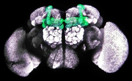
Mushroom bodies in Drosophila brain
|
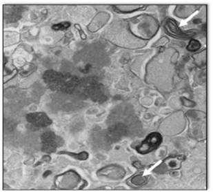
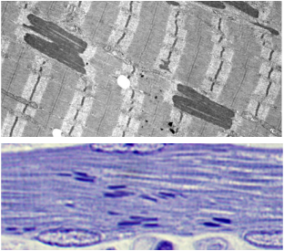
Myelinoid bodies (arrowed)
|
Myelinoid bodies - Pleomorphic structures such as lysosomes that contain a myelin figure. Also known as myelinosomes (local myelin out-foldings) they are often the result of early myelin damage. Myelinoid bodies have been associated with neuroendocrine cells (particularly of the prostatourethral region) and disorders such as renal glomerulonephritis. They are also associated with curvilinear bodies in hydroxychloroquine cardiotoxicity (also see Curvilinear bodies)
Myeloid bodies - Distinct lamellar regions of the smooth endoplasmic reticulum found within the retinal pigment epithelial cells in a number of lower vertebrates. In humans, they are associated with the lysosomal storage disorder phospholipidosis. Although this disorder may be drug induced, these bodies have also been identified in renal tubules following treatment with gentamicin
Myeloid bodies - Distinct lamellar regions of the smooth endoplasmic reticulum found within the retinal pigment epithelial cells in a number of lower vertebrates. In humans, they are associated with the lysosomal storage disorder phospholipidosis. Although this disorder may be drug induced, these bodies have also been identified in renal tubules following treatment with gentamicin
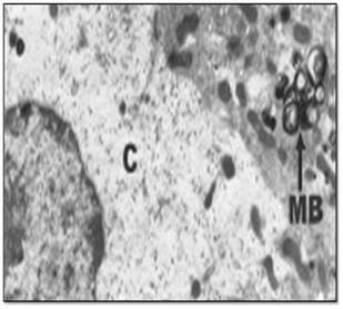
Myeloid bodies (MB) in the cytoplasm (C)
|
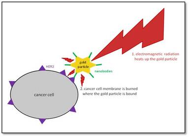
Nanobody
|
Nanobodies - These are single-domain antibodies derived from the immunoglobulins of camels and llamas (Camelidae). They were originally developed following the discovery that the antibodies consisted of heavy chains but lacked light chains. Nanobodies, which are able to bind tumour antigens such as Her2, are coupled to gold nanoparticles that absorb light energy and create heat in order to kill cancer cells
Nasal swell bodies - Also known as the septal turbinate, the nasal swell body is the widened region of the septum that is located in the nose. The body consists of cartilage and bone overlying a thickened mucosa and is important in regulating the airflow through the nasal valve. The bodies are generally thicker in patients with allergic rhinitis
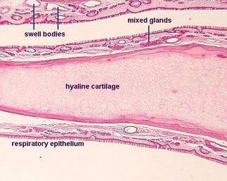
Nasal swell bodies
|
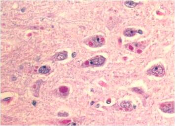
Negri bodies
|
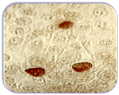
Negri bodies - Round or oval cytoplasmic inclusions in nerve cells of the brain in patients infected with rabies. Staining with Giemsa can differentiate Negri bodies from other intracellular inclusions by staining magenta with dark-blue granules. They are sometimes referred to as Babes-Negri bodies
Neill-Mooser bodies - These are large mononuclear cells filled with Ricketssia typhi (Gram negative bacteria) in patients with endemic typhus fever. Also known as Mooser bodies, the term refers to the bacteria that are found in the epithelium and exudate of the tunica vaginalis in infected patients
Neill-Mooser bodies - These are large mononuclear cells filled with Ricketssia typhi (Gram negative bacteria) in patients with endemic typhus fever. Also known as Mooser bodies, the term refers to the bacteria that are found in the epithelium and exudate of the tunica vaginalis in infected patients
Neill-Mooser bodies
|
Nemaline bodies
|
Nemaline bodies - These are abnormal thread-like rods found in skeletal muscle in patients with nemaline myopathy. Nemaline or rod body myopathies are a group of conditions which fall under the category of congenital myopathies
Neuroepithelial bodies - Small, discrete collections of pulmonary neuroendocrine cells that is sensitive to hypoxia. Structural changes in them have been seen in humans living at high altitude. Innervated clusters of these bodies are believed to function as pulmonary airway chemoreceptors sensitive to low oxygen levels. However, data is vague on the effect high altitude has on these pulmonary cells
Neuroepithelial bodies - Small, discrete collections of pulmonary neuroendocrine cells that is sensitive to hypoxia. Structural changes in them have been seen in humans living at high altitude. Innervated clusters of these bodies are believed to function as pulmonary airway chemoreceptors sensitive to low oxygen levels. However, data is vague on the effect high altitude has on these pulmonary cells
Neuroepithelial bodies
|
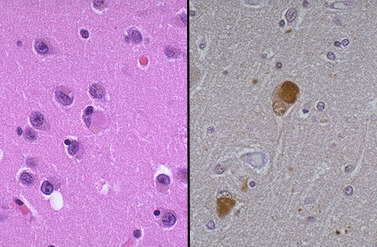
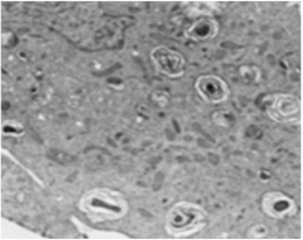
Nissl bodies
|
Nissl bodies - Large granular basophilic bodies found in the cytoplasm of neurones, also known as Chromophilous bodies. They are composed of rough endoplasmic reticulum and ribosomes and are involved with protein synthesis. They are rich in RNA and stain strongly with basic dyes. The image above is stained with Luxol Fast Blue and Cresyl fast Violet and shows purple Nissl bodies with myelin stained blue. They are synonymous with Tigroid bodies which are defined as any one of the large granular structures in the cytoplasm of nerve cells that stains with basic dyes and contains ribonucleoprotein
Nuclear bodies - Nuclear bodies are a collective term for a variety of discrete substructures that are found in eukaryotic nuclei. The absence of membranes around them allows their components to exchange more freely with the surrounding nucleoplasm. Some of the bodies are involved in RNA metabolic processes (see separate entry for Cajal body), while others are associated with a variety of processes such as transcription, protein modification and apoptosis. Location of nuclear bodies such as the Cajal body, Cleavage body, Histone locus body, PcG (polycomb group) body, PML (promyelocytic leukemia) and SAM 68 bodies can be seen in the Figures below. Many other nuclear bodies such as the HAP (heterogeneous ribonucleo-associated protein), Orphan and Nuclear stress bodies have also been documented. However, it is beyond the scope of this website to describe in detail the structure and function of all nuclear bodies and a complete descriptive list of them is best obtained from alternative sources
Nuclear bodies - Nuclear bodies are a collective term for a variety of discrete substructures that are found in eukaryotic nuclei. The absence of membranes around them allows their components to exchange more freely with the surrounding nucleoplasm. Some of the bodies are involved in RNA metabolic processes (see separate entry for Cajal body), while others are associated with a variety of processes such as transcription, protein modification and apoptosis. Location of nuclear bodies such as the Cajal body, Cleavage body, Histone locus body, PcG (polycomb group) body, PML (promyelocytic leukemia) and SAM 68 bodies can be seen in the Figures below. Many other nuclear bodies such as the HAP (heterogeneous ribonucleo-associated protein), Orphan and Nuclear stress bodies have also been documented. However, it is beyond the scope of this website to describe in detail the structure and function of all nuclear bodies and a complete descriptive list of them is best obtained from alternative sources
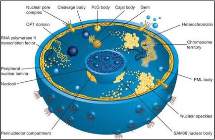
Nuclear bodies - 1
|
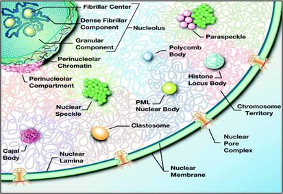
Nuclear bodies - 2
|
Nuclear stress bodies - see Nuclear bodies
Nucleolar accessory bodies – see Cajal bodies