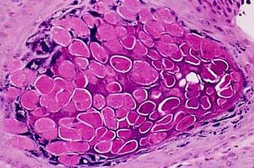
Farber bodies - These are curvilinear, tubular structures found in cytoplasmic vacuoles of tissues in patients with Farber’s disease, a genetically determined disorder of lipid metabolism. The disease is associated with deficiency of lysosomal acid ceramidase and accumulation of ceramide in the lysosomes. In adults, elevated levels of ceramide have been found in subcutaneous nodules in the liver, kidney, lungs and brain. Children who have the classic form of Farber's disease develop symptoms within the first few weeks of life which are characterized by joint deformity and the presence of subcutaneous nodules (see image)
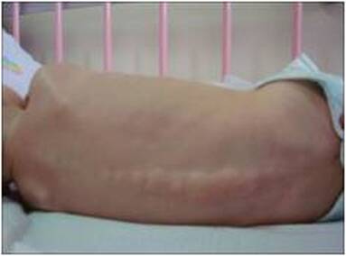
Subcutaneous nodules along spine in Farber's disease
|
Fardeau-Engel bodies
|
Fardeau-Engel bodies - These are polygonal structures found in the cytoplasm of Schwann cells. Located in unmyelinated nerve fibres, they contain crystalline shapes and are found in certain conditions such as Refsum disease (peripheral neuropathy). In striated muscle, these bodies may sometimes be mistaken for abnormal mitochondria (mitochondriopathy)
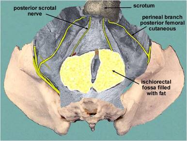
Fat bodies - These are nutritional reservoirs of fatty tissue within the ischioanal and ischiorectal fossa and found mainly in amphibians and insects. Also check out Orbital fat and Oval fat bodies
Fat bodies - These are nutritional reservoirs of fatty tissue within the ischioanal and ischiorectal fossa and found mainly in amphibians and insects. Also check out Orbital fat and Oval fat bodies
Fat bodies
Ferruginous bodies - see Asbestos bodies
Fessas bodies - These are intracellular inclusion bodies found in peripheral blood smears of patients suffering from thalassemias. These are disorders of decreased synthesis and defective production of normal haemoglobin. It is thought that Fessas bodies are aggregates of the alpha chains of haemoglobin
Fessas bodies - These are intracellular inclusion bodies found in peripheral blood smears of patients suffering from thalassemias. These are disorders of decreased synthesis and defective production of normal haemoglobin. It is thought that Fessas bodies are aggregates of the alpha chains of haemoglobin
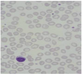
Fessas bodies
|
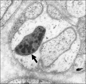
Fibrin body in alveolar capillary
|
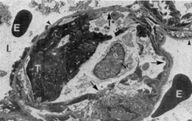
Fibrin bodies - Movable or adherent, round, sharply demarcated opacities near the base of the pleural cavity. Also termed pleural fibrin balls, they may occur secondary to pleural effusion and pneumothorax. They are formed by blood fibrin separating out from a blood clot. The image shows a fibrin body or thrombus (T) in a capillary and red blood cells (E) in an alveolar lumen (L)
Fibrous bodies – These are round, eosinophilic, perinuclear structures that occur exclusively in the growth hormone and acidophil cells of the pituitary. They are strongly reactive for low molecular weight cytokeratins and have also been termed Keratin bodies. They are located in the Golgi region and are reliable markers in the differential diagnosis of pituitary adenomas
Fibrous bodies – These are round, eosinophilic, perinuclear structures that occur exclusively in the growth hormone and acidophil cells of the pituitary. They are strongly reactive for low molecular weight cytokeratins and have also been termed Keratin bodies. They are located in the Golgi region and are reliable markers in the differential diagnosis of pituitary adenomas
Fibrous bodies (circled)
|
Fingerprint bodies
|
Fingerprint bodies - These bodies are found in the congenital benign muscle disorder known as fingerprint body myopathy. The disease is characterised by low muscle tone and weakness with fingerprint bodies located at the periphery of the muscle fibres
Flemming bodies - Round, deeply staining bodies found in the germinal centres of lymph follicles and in the cytoplasm of phagocytes. They are also known as midbodies or Tingible Korper bodies. They are present towards the end of the complete separation of dividing cells and are numerous in the vicinity of mitotic figures
Flemming bodies - Round, deeply staining bodies found in the germinal centres of lymph follicles and in the cytoplasm of phagocytes. They are also known as midbodies or Tingible Korper bodies. They are present towards the end of the complete separation of dividing cells and are numerous in the vicinity of mitotic figures
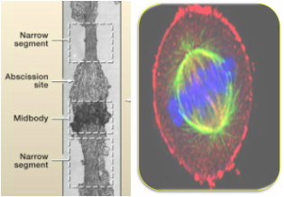
Flemming (mid) body
|
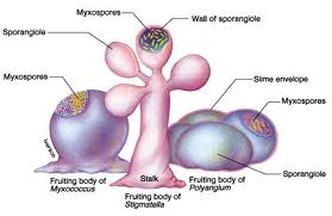
Fruiting bodies
|
Fruiting bodies - These are specialized macroscopic reproductive structures that are produced by certain types of fungi and bacteria (such as myxobacteria), some of which are pathogenic in fish and other animals
Fuchsin bodies – see Russell bodies
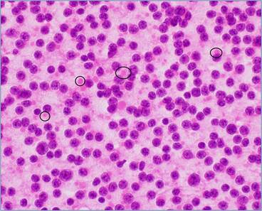
Gall bodies - Small round vacuoles found in lymphocytes that correspond to lysosomes filled with carotenoids and lipid. Carotenoid levels are age-dependent, decreasing in the elderly
Fuchsin bodies – see Russell bodies
Gall bodies - Small round vacuoles found in lymphocytes that correspond to lysosomes filled with carotenoids and lipid. Carotenoid levels are age-dependent, decreasing in the elderly
Gall bodies
|
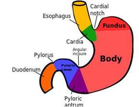
Gastric body
|
Gastric bodies - Body of the stomach between the pylorus and the fundus, also known as the corpus gastricum
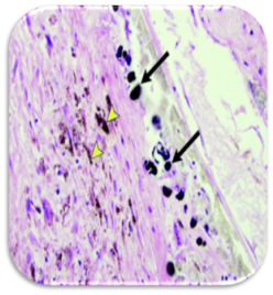
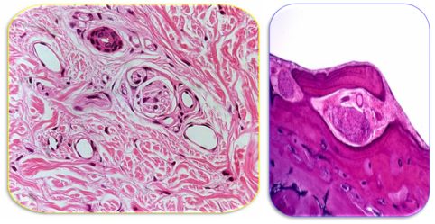
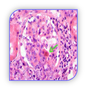
Gamna-Favre bodies - Basophilic inclusion bodies located in the cytoplasm of endothelial cells. They are found in lymphogranuloma venereum (LGV - see image), a sexually transmitted disease caused by Chlamydia trachomatis. The disease is also called Durand-Nicholas-Favre disease, lymphopathia venereum, lymphogranuloma inguinale, tropical bubo, and poradenitis inguinales. Gamna-Favre bodies are relatively large and probably composed of degenerated nuclear material
LGV - see Gamna-Favre bodies
|
Gamna-Gandy bodies
|
Gamna-Gandy bodies - Nodules that are secondary to the accumulation of haemosiderin. They are found in the liver and spleen of patients with cirrhosis. Haemosiderin can also complex with calcium in splenic scars
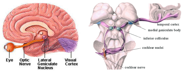
Geniculate bodies - These are four small oval masses that protrude slightly from the underside of the thalamus and function as synaptic centres on the way to the cerebral cortex. The lateral geniculate body is associated with sight (left hand image) while the medial geniculate body is concerned with hearing (right hand image)
Geniculate bodies
|
Giannuzzi bodies
|
Giannuzzi bodies - Crescent-shaped patches of serous cells surrounding the tubules in mucous glands. They are formed by multiple cells that are pushed to the blind ends of the terminal portions of the glands. They are also known as Giannuzzi’s crescents, serous demilunes or semilunar bodies (also see Crescent bodies)
Glass bodies - see Crescent bodies
Globoid bodies - Small tubular structures or inclusions found in Schwann cells. They are found in Krabbe’s disease, a neurodegenerative disorder of infancy. The disease is generally characterized by the onset of symptoms before the age of 6 months with death usually occurring by the end of the first year. Clinically, the child fails to thrive, developing seizures, deafness, blindness, paralysis and marked mental deficiency
Globoid bodies
|
Glomus bodies
|
Glomus bodies - Ball-like swellings of the skin containing nerve fibres. They help to regulate the oxygen pressure in the middle ear and respond to change in body temperature and blood pressure. Glomus tumours are benign lesions of the middle ear which arise from glomus bodies
Golgi bodies - Golgi bodies consist of flattened membrane sacs in the cytoplasm of cells. They are the centre for glycoprotein metabolism and are involved in cell division. Known also as the Golgi apparatus or Golgi vesicles, they gather protein and lipid for dispatch to specific locations in the cell
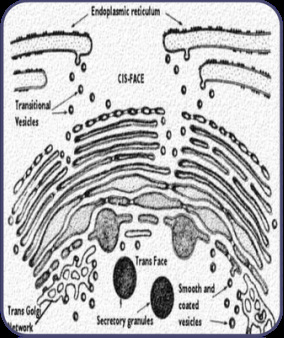
Golgi bodies
|
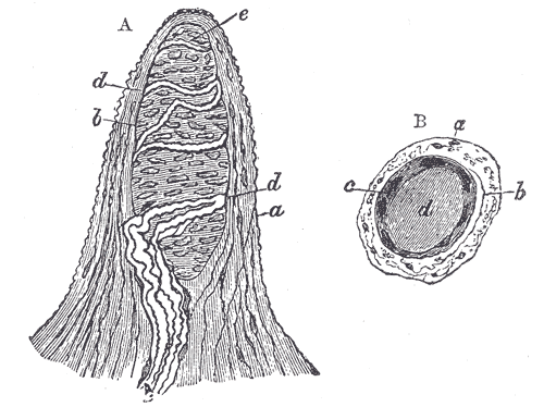
Golgi-Mazzoni bodies -Diagram of a fingertip - Figure A: a. Cortical layer. b. Tactile corpuscle. c. Small nerve of the papilla. d. Two nerve fibres with spiral coils around corpuscle. e. Termination of fibres. Figure B: a. Cortical layer. b. Nerve fibre. c. Outer layer of the tactile body. d. Clear interior substance
|
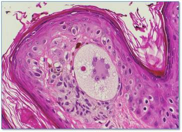
Golgi-Mazzoni bodies - Encapsulated sensory nerve endings found in the subcutaneous tissue of the fingertips (see image above). They differ from pacinian corpuscles by having thinner capsules, possess fewer lamellae and contain fibres that are more extensively branched. Also see Vater-Pacini bodies
Gottlieb bodies - Eosinophilic inclusion bodies found in the vesicular cytoplasm of multinucleated melanocytes. They are associated with benign naevi, particularly those of long duration
Grynfellt bodies - see Buscaino bodies
Grynfellt bodies - see Buscaino bodies
Gottlieb body
Guarnieri bodies - Round or oval cytoplasmic inclusion bodies found in patients with smallpox. They are slightly basophilic or acidophilic and lie close to the nuclei
Guarnieri bodies
|
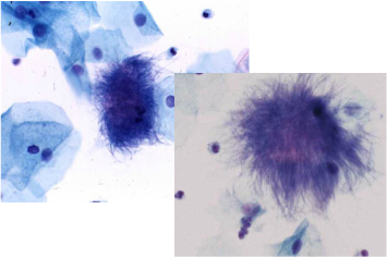
Gupta bodies
|
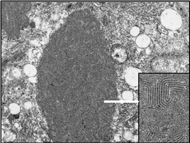
Gupta bodies - Aggregates of filamentous, branching material often with a woolly appearance. The periphery of them may contain swollen filaments with club formation. Often called ‘dust bunnies’, they can be seen in cervical smears in women infected with Actinomyces israelii as a result of long term use of intrauterine contraceptive devices
GW bodies - Cytoplasmic structures that were first considered as both storage centres and degradation sites for a specific subset of messenger RNA. They contain glycine (G) and tryptophan (W) and are consequently termed GW bodies. Known to vary in size and number throughout the cell cycle, they were initially identified through the use of an autoimmune serum that targeted the marker protein GW182. GW bodies are one of the most identified cellular targets of autoantibodies in primary biliary cirrhosis. These bodies are sometimes referred to as mammalian processing (P) bodies (see separate entry for Processing bodies)
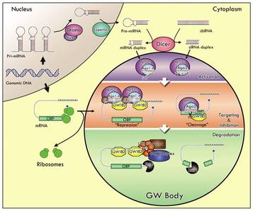
Location of the GW body
|
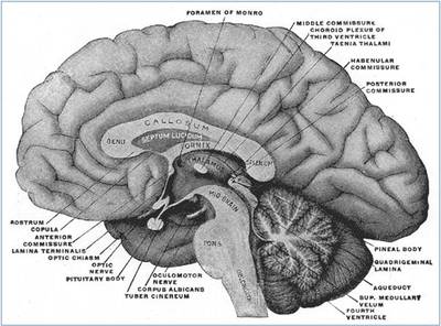
Habenular body near the thalamus towards the base of the brain
|
Habenular bodies - These comprise small groups of nuclei that are situated at the posterior end of the thalamus at the region where the pineal gland attaches to the brain
Haematoxylin bodies - Dense, homogeneous particles consisting of denatured nuclear material from injured cells. These are found in patients with systemic lupus erythematosus (SLE) and are found more frequently in the renal glomeruli. Lymphocytes that ingest such particles are termed LE cells
Haematoxylin bodies
|
Halberstaedter-von Prowazek bodies
|
Halberstaedter-von Prowazek bodies - Intracytoplasmic inclusions that vary in size from minute (elementary bodies) to large (reticulate or initial bodies). They represent the causal agent of trachoma (chlamydia trachomatis) and are sometimes known as Trachoma bodies. The disease is spread by aerosol or contact although flies may help transfer ocular infections in endemic areas. See Elementary and Reticulate bodies and also Coccoid X bodies for psittacosis, another type of chlamydial infection
Hamazaki-Wesenberg bodies - Variably-sized ovoid inclusions that stain positive with PAS and methenamine silver. Also known as Yellow-Brown bodies, they are found in lymph nodes and are thought to be related to the lipofuscins. Because they sometimes appear as budding forms, they may be misinterpreted as fungi. Hamazaki-Wesenberg bodies are found in granulomatous diseases such as sarcoid.
HAP (Heterogenous ribonucleo-associated protein) bodies - see Nuclear bodies
Hamazaki-Wesenberg bodies
|
Harting bodies
|
Harting bodies - Minute globular bodies formed during calcification by chemical union of calcium particles and the albuminous matter of cells. They are found in cerebral capillaries and are termed calcospherites
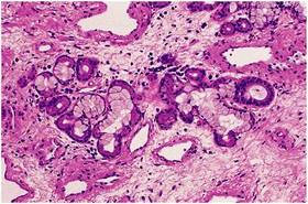
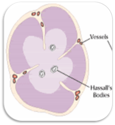
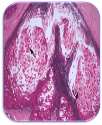
Hassall bodies - Small spherical bodies of keratinised and usually squamous epithelial cells arranged in a concentric pattern around clusters of degenerating lymphocytes, eosinophils, and macrophages. Also known as Thymus and Virchow-Hassall bodies, they can be found in the medulla of the thymus at the fifteenth week of fetal development
Hassall bodies - Small spherical bodies of keratinised and usually squamous epithelial cells arranged in a concentric pattern around clusters of degenerating lymphocytes, eosinophils, and macrophages. Also known as Thymus and Virchow-Hassall bodies, they can be found in the medulla of the thymus at the fifteenth week of fetal development
Hassall bodies
|
Hassall-Henle bodies
|
Hassall-Henle bodies - Small hyaline excrescences found on the posterior surface of Descemet's membrane at the periphery of the cornea. They contain numerous cracks and fissures filled with extrusions of corneal epithelium. These bodies are found in large quantities in degenerative disease and chronic inflammation and are probably associated with the aging process
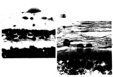
Hectoid bodies - These are associated with sickle cell anaemia, a genetic blood disorder. The disease is characterized by red blood cells assuming an abnormal sickle shape as a result of a mutation in the haemoglobin gene (see image below). However, it is not clear what the characteristics of hectoid bodies are
Hectoid bodies - These are associated with sickle cell anaemia, a genetic blood disorder. The disease is characterized by red blood cells assuming an abnormal sickle shape as a result of a mutation in the haemoglobin gene (see image below). However, it is not clear what the characteristics of hectoid bodies are
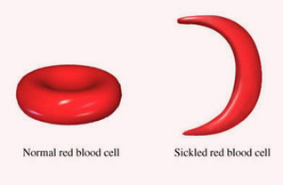
Sickled red blood cell
|
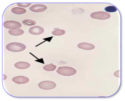
Heinz bodies
|
Heinz bodies - These are small rounded distensions that deform red blood cells. They are found in glucose-6-phosphate dehydrogenase deficiencies but also found in congenital haemolytic anaemias and in premature infants. Also known as Ehrlich or Heinz-Ehrlich bodies, they occur as a result of damage to the haemoglobin molecule. They are best seen when blood films are stained with dyes such as crystal violet or methylene blue
Heinz-Ehrlich bodies – see Heinz bodies
Hemispherical bodies - These are found in the eye of patients with glaucoma and traumatic retinal detachment. They have been associated with eyes fixed in Zenker’s fixative but not with those fixed in formalin
Heinz-Ehrlich bodies – see Heinz bodies
Hemispherical bodies - These are found in the eye of patients with glaucoma and traumatic retinal detachment. They have been associated with eyes fixed in Zenker’s fixative but not with those fixed in formalin
Hemispherical bodies in the eye
|
Henderson-Patterson bodies
|
Henderson-Patterson bodies - Eosinophilic, cytoplasmic inclusions found in the cells of the stratum spinosum and corneum. They are seen in the disease molluscum contagiosum, a non-cancerous viral infection of the skin caused by the poxvirus. The bodies comprise a membrane sac containing numerous viral particles. Viral infection leads to the development of the molluscum lesions on the skin but does not affect any internal organs. Also known as Molluscum bodies
Hensen bodies - These bodies are found in the outer hair cells of the inner ear and are responsible for the recycling of membrane proteins in the hair cells. These bodies are often the starting point for the medical treatment of hearing impairments caused by noise
Hensen bodies - These bodies are found in the outer hair cells of the inner ear and are responsible for the recycling of membrane proteins in the hair cells. These bodies are often the starting point for the medical treatment of hearing impairments caused by noise
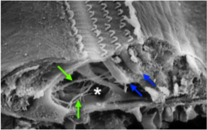
Hensen bodies (blue arrows); the star indicates the tunnel of Corti where nerve fibres are crossing and the green arrows are afferent nerve fibres
|
Herring bodies
|
Herring bodies - Large eosinophilic masses of neurosecretory granules located in the posterior lobe (pars nervosa) of the pituitary at dilatations along the axons and their endings. The posterior pituitary releases anti-diuretic hormone (ADH) and oxytocin from these bodies. ADH is also known as vasopressin, a hormone that acts on the kidneys
Highmore bodies - Mass of fibrous tissue continuous with the tunica albuginea that project into the testis. Highmore bodies are known as mediastinum testis
Highmore bodies - Mass of fibrous tissue continuous with the tunica albuginea that project into the testis. Highmore bodies are known as mediastinum testis
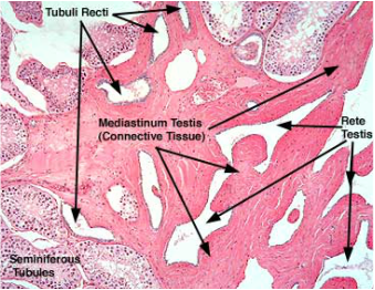
Highmore bodies (mediastinum testis)
|
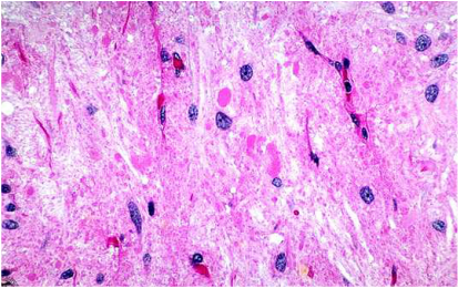
Hirano bodies
|
Hirano bodies - These are intracellular, eosinophilic and often rod-shaped structures that are found in nerve cells. They occur in neurodegenerative diseases such as Alzheimer's and Creutzfeldt-Jacob disease and are composed of microfilament-associated proteins such as actin and vinculin
Histone locus bodies - see Nuclear bodies
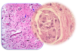
Howell-Jolly bodies - Spherical, blue-black inclusions of red blood cells in smears stained with Wright stain. They are seen in severe haemolytic anaemias, in patients with dysfunctional spleens and after splenectomy. The bodies are nuclear fragments of condensed DNA that are normally removed by the spleen. The host red cell usually contains only a single body which appears as a perfectly round inclusion
Histone locus bodies - see Nuclear bodies
Howell-Jolly bodies - Spherical, blue-black inclusions of red blood cells in smears stained with Wright stain. They are seen in severe haemolytic anaemias, in patients with dysfunctional spleens and after splenectomy. The bodies are nuclear fragments of condensed DNA that are normally removed by the spleen. The host red cell usually contains only a single body which appears as a perfectly round inclusion
Howell-Jolly bodies
|
Hyaline bodies
|
Hyaline bodies - These are linear or filamentous granules that are found in a variety of conditions such as myopathy and hepatocellular carcinoma. They are also found in optic nerve head drusen where hyaline bodies are often retained (also see Drusen bodies)
Hyaloid bodies - see Vitreous bodies
Infrapatellar fat bodies - The infrapatellar fat bodies known as Hoffa’s fat pads occupy the area between the patellar ligament and the synovial fold of the knee joint. They cushion the patella in the event of a direct blow to the front of the knee
Hyaloid bodies - see Vitreous bodies
Infrapatellar fat bodies - The infrapatellar fat bodies known as Hoffa’s fat pads occupy the area between the patellar ligament and the synovial fold of the knee joint. They cushion the patella in the event of a direct blow to the front of the knee
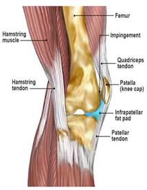
Infrapatellar fat body
|
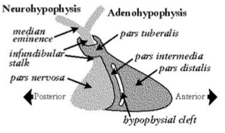
Infundibular body
|
Infundibular bodies - The infundibular body or stalk is the funnel-shaped pituitary stalk that connect the hypothalamus of the brain with the posterior pituitary
Initial bodies - see Reticulate bodies
Interrenal bodies - Elongated tissue situated between the kidneys of certain fishes which correspond with the adrenal medulla of mammals
Initial bodies - see Reticulate bodies
Interrenal bodies - Elongated tissue situated between the kidneys of certain fishes which correspond with the adrenal medulla of mammals
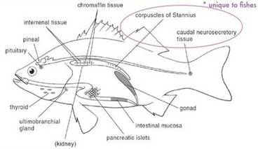
Interrenal body or tissue
|
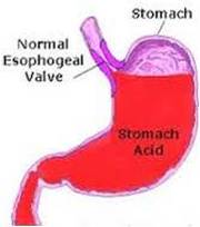
Gastric juices - see Jaworski bodies
|
Jaworski bodies - Spiral mucous bodies or corpuscles seen in the secretion of the stomach in hyperchlorhydria, an excess of hydrochloric acid in gastric juice that is often characteristic of some pathological states
Joest bodies – see Joest-Degen bodies
Joest-Degen bodies - Intranuclear and eosinophilic inclusions found in nerve cells of warm-blooded animals with Borna disease. This is an infectious neurological syndrome caused by the Borna disease virus, an RNA virus. The disease causes abnormal behaviour and fatality in infected animals
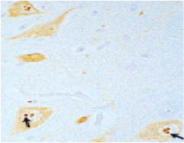
Joest-Degen bodies
|
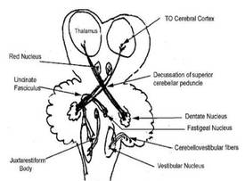
Juxtarestiform body
|
Juxtarestiform bodies – These are a subdivision of the inferior cerebellar peduncle, (which comprise both the juxtarestiform and restiform bodies) and carry both afferent and efferent nerve fibres. The bodies have a role in coordinating balance and eye movements (also see Restiform bodies)